Apple's QuickTime Plugin is required to view these movies. If you do not already have QuickTime installed, you can obtain the latest version at http://www.apple.com/quicktime/download/.
See
Article: [Science & Technology: Technology Roundup Digital Video Providing
broad accesss to Tissue Banks]
Cell Identification Movie (1.0 MB)
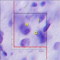
This through-focus movie of the dorsal lateral geniculate nucleus demonstrates our procedures for cell counting and identification. This movie was created from a portion of the hi-res through-focus movie below. Abbreviations:
- E: Endothelial Cell
- G: Glial Cell
- I: Interneuron
- N: Principal Neuron
- U: Unclassified Cell
|
|
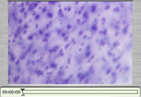
High Resolution Through-Focus Movie
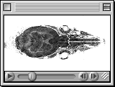
A through-focus series of the thalamus (36 frames through the dorsal lateral geniculate nucleus) cut in the coronal plane. This is just a very small central piece of the whole field. The material is from a BXD5 female (celloidin ID 241, slide A, section 23 left, case ID 072397A). The tissue was imaged with a 40X oil objective (Olympus UVFL NA 1.3) used in combination with DIC optics. Pixel resolution in the XY plane is 0.09 µm/pixel (optic resolution approximatley 0.3 µm and the field is 275 x 183 µm. The Z axis step averages 1.0 µm. The image stack was acquired manually over a 5 minute interval using the PhaseOne digital camera back (3056 by 2032 pixels, 8-bit color). You will need a QuickTime plug-in to step through the sections. Make sure your browser has plenty of memory. Two smaller preview movies are also available:
- Small zoomed-in sample from the center of the full field that demonstrates the maximum resolution of the large image stack (0.4 MB).
- Smaller down-sampled version of the original that shows the entire field. (0.9 MB).
- High Resolution Through-Focus Movie. CAUTION: 23.8 MB! Download this file to disk and then play locally using the QuickTime player. This is the whole field at its original capture resolution. You will need a large monitor to view the field at even a 50% reduction. The best way to view the z axis stack is to select "full screen" in the QuickTime Player after you have opened the file. For improved performance, assign QuickTime ample memory.
|
|
|
Mouse Brain Fly-Through Movie (0.9 MB): Sections of a brain cut in the horizontal plane (C57BL/6J). Resolution is 24.5 µm/pixel. Sections are spaced 150 µm apart. You will need a QuickTime plug-in to step through the sections. Make sure your browser has plenty of memory. A smaller (0.2 MB) preview movie is also available:
|
|
|
Mouse Brain Stack Movie (1.0 MB): In this version, the sections stack on top of each other from ventral to dorsal. A smaller (0.2 MB) preview movie is also available.
|
|
|
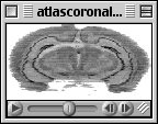
3D Atlas of a Mouse Brain (2.2 MB): We stained and photographed every horizontal section of case 020 from the MBL (a 129/SvJ, 39-day-old male with a 386 mg brain). Dr. Nissanov and colleagues at Drexel/MCP Hahnemann University used these images to construct a complete 3D digital representation of the brain at a resolution of approximately 30 µm in all axes (i.e., an isotropic 3D atlas). This 3D model is like a super-high resolution MRI and can be digitally sectioned in any axis. Here we show a fly-through of the brain in the frontal axis, at right angle to the sectioning plane. High-resolution color images of whole mouse brains are also available.
|
|
|
High Magnification Movie (0.4 MB): A through-focus series of the hilus region of the dentate gyrus. This movie illustrates some features of the direct 3-dimensional counting method. Step through the movie frame-by-frame. Each frame is separated by 1 µm in the z axis. The scale bar is divided into 2-µm units.
|
|
|
MRI Fly-Through of a Mouse Head and Brain in the Frontal Plane. This QuickTime movie was generated from a 62.5 micron/pixel isotrophic scan of a female BXD5-Ty mouse by E. Ahrens, R. Jacobs, and R. W. Williams (1996 unpublished) taken with the 12 Tesla Caltech system. Each frame is 200 by 200 pixels.
|
|